
What Is Breast Thermography?
Thermography or Medical infrared imaging (MII) is a safe, non-invasive medical screening procedure that detects and records infrared heat patterns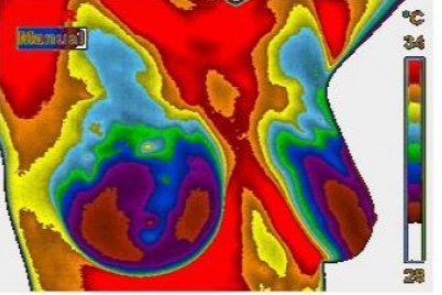 or markers from the breast, such as increased blood vessel or capillary formation, which can aid in the early detection of abnormal changes in breast tissue.
While mammography, ultrasound, MRI, and other structural imaging tools rely primarily on finding the physical tumor, MII detects the heat produced by increased blood vessel circulation and metabolic changes associated with a tumor’s genesis and growth.
By detecting minute variations in normal blood vessel activity, infrared imaging may find thermal signs suggesting pre-cancerous breast tissue or the presence of an early tumor that is not yet large enough to be detected by physical examination, mammography, or other types of structural imaging.
Certain types of cancers will not be detected by mammography for various reasons, but some of these cancers will be discovered by MII.
Difficulties in reading mammograms can occur in women who are on hormone replacement, nursing or have fibrocystic, large, dense, or enhanced breasts. These types of breast differences do not cause difficulties in reading digital infrared scans.
MII AS A RISK MARKER FOR BREAST CANCER
Studies show that an abnormal infrared image is the single most important marker of high risk for developing breast cancer, 10 times more significant than a family history of the disease. Consequently, in patients with a persistent abnormal thermogram, the examination results become a marker of higher future cancer risk. Depending upon certain factors, re-examinations are performed at appropriate intervals to monitor the breasts. This gives a woman time to take a proactive approach to improve her breast health. By maintaining close monitoring of her breast health with infrared imaging, self breast exams, clinical examinations, and other tests, a woman has a much better chance of detecting cancer at its earliest stage and preventing invasive tumor growth.
Just as unique as a fingerprint, each patient has a particular infrared map of their breasts. Any modification of this infrared map on serial imaging (images taken over months to years) may constitute an early sign of an abnormality.
By carefully examining changes in the temperature and blood vessels of the breasts, signs of possible cancer or precancerous cell growth may be detected years prior to being discovered using any other procedure.